Introduction
Since orthopedic surgeons began using a tiny TV camera called an arthroscope to diagnose and treat shoulder problems, they have discovered several conditions that no one knew existed. One of these conditions is an injury to a small structure in the shoulder called the labrum. A labral tear can cause pain and a catching sensation in the shoulder. Labral tears can be very difficult to diagnose.
This guide will help you understand
- Where and what the labrum is
- What tests will Dr. Kiritsis run to diagnose the problem
- What you can do to relieve your pain
Anatomy
The shoulder is made up of three bones: the scapula (shoulder blade), the humerus (upper arm bone), and the clavicle (collarbone).
A part of the scapula, called the glenoid, makes up the socket of the shoulder. The glenoid is very shallow and flat. The labrum is a rim of soft tissue that makes the socket more like a cup. The labrum turns the flat surface of the glenoid into a deeper socket that molds to fit the head of the humerus.
The rotator cuff connects the humerus to the scapula. The rotator cuff is formed by the tendons of four muscles: the supraspinatus, infraspinatus, teres minor, and subscapularis.
Tendons attach muscles to bones. Muscles move the bones by pulling on the tendons. The rotator cuff helps raise and rotate the arm. As the arm is raised, the rotator cuff also keeps the humerus tightly in the glenoid of the scapula.

The soft labral tissue can be caught between the glenoid and the humerus. When this happens, the labrum may start to tear. If the tear gets worse, it may become a flap of tissue that can move in and out of the joint, getting caught between the head of the humerus and the glenoid.
The flap can cause pain and catching when you move your shoulder. Several tendons and ligaments attach to the labrum which helps maintain the stability of the shoulder. So when the labrum tears, the shoulder often becomes much less stable.
Related Document: A Patient’s Guide to Shoulder Anatomy
Causes
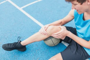
Labral tears are often caused by a direct injury to the shoulder, such as falling on an outstretched hand. The labrum can also become torn from the wear and tear of activity, a condition called overuse. Instability in the shoulder may lead to repetitive stress and eventual tearing of the labrum. An extremely unstable shoulder may slip or dislocate. This will cause the labrum to tear.
Related Document: A Patient’s Guide to Shoulder Instability
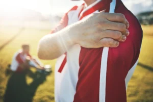
The biceps tendon attaches to the front part of the labrum. The biceps is the large muscle on the front of your upper arm. Certain sports can cause injuries to the labrum when the biceps tendon pulls sharply against the front of the labrum. Baseball pitchers and overhead athletes are prone to labral tears as contact athletes like football and lacrosse players.
Related Document: A Patient’s Guide to Biceps Tendonitis
Symptoms
The main symptom caused by a labral tear is a sharp pop or catching sensation in the shoulder during certain movements. This may be followed by a vague aching for several hours. At other times, the tear may not cause any pain.
Shoulder instability from a damaged labrum may cause the shoulder to feel loose, as though it slips with certain movements. It is important to remember that labral tears are very common in athletes even when they don’t have any shoulder pain. Forty percent of professional baseball players with no shoulder pain has a labral tear.
Diagnosis
Dr. Kiritsis may suspect a labral tear based on your medical history. You will be asked questions about your pain and past injuries to your shoulder that may suggest labral damage.
In the physical examination, there are several shoulder movements that can bring on the symptoms. You may feel a catching sensation as your arm is raised, and there may be a pain when the arm is held overhead. If your arm is held in front of your body, with the palm of the hand facing downward, you may feel pain when Dr. Kiritsis tries to push down your arm.
Labral tears are difficult to see, even in a magnetic resonance imaging (MRI) scan. An MRI scan is a special imaging test that uses magnetic waves to show the tissues of the shoulder in slices. The MRI scan shows soft tissues such as tendons and ligaments as well as bones.
Labral tears may be seen using computed tomography (CT) scan and a special dye. A CT scan is an older test that uses computer-enhanced X-rays to show slices of the shoulder. The soft tissues do not show up in a CT scan, but the special dye does. The dye shows the outline of the labrum. If there is a tear, the dye may leak into it and show up on the CT scan.
However, MRI and CT scans are not very accurate in detecting labral tears. Confirming the diagnosis of a labral tear can be extremely difficult. Dr. Kiritsis may need to look into your shoulder using an arthroscope. The arthroscope is a small TV camera that is inserted into the shoulder joint through a very small incision. Dr. Kiritsis can then see pictures of the joint on a TV screen which allows him to look directly at the labrum to see if it is torn.
Nonsurgical Treatment
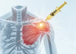
Dr. Kiritsis’ first goal will be to control your pain and inflammation. Initial treatment for pain control is usually rest and anti-inflammatory medication, such as aspirin or ibuprofen. He may suggest a cortisone injection if you have trouble getting your pain under control. Cortisone is a strong anti-inflammatory medication. It can provide good relief, although its effects are temporary. We try to avoid injecting the shoulders of our young athletes.
Dr. Kiritsis will have a physical or occupational therapist direct your rehabilitation program. Your first therapy treatments will try to ease pain and inflammation by using such treatments as heat or ice. Hands-on treatment and various types of exercises are used to improve the range of motion in your shoulder and the nearby joints and muscles.
Later, you will do strengthening exercises to improve the strength and control of the rotator cuff and shoulder blade muscles. Your therapist will help you retrain these muscles to keep the ball of the humerus centered in the glenoid. This will improve the stability of your shoulder and help it move smoothly during all your activities.
You may need therapy treatments for four to six weeks. Most patients are able to get back to their activities with full use of their arms within this amount of time.
Surgery
If your symptoms persist, you may need surgery. Dr. Kiritsis is able to provide his patients with the latest arthroscopic techniques even in the most demanding cases.
Labral Debridement

The arthroscope is commonly used to treat many labral tears. If the tear is small and is mostly getting caught as you move the shoulder, simply removing the frayed edges and any loose parts of the labrum may get rid of your symptoms. This is called labral debridement.
Labral Repair

If the tear is larger, the shoulder may also be unstable. If this is the case, the labral tear may need to be repaired, rather than simply removed. Several new techniques allow Dr. Kiritsis to place anchors into the bone around the shoulder joint and reattach the labrum using the arthroscope.
There are many variations of these anchors, but most are drilled into the bone and have sutures (stitches) attached that are then used to tie the labrum down to the bone and enable the labrum to heal back in the appropriate position.
Open Procedure
Open procedures are rarely used today for the repair of labral tears. The arthroscopic techniques have become increasingly refined and are today the preferred method of treatment.
Nonsurgical Rehabilitation
Even nonsurgical treatment requires a rehabilitation program. The goal of therapy will be to strengthen the rotator cuff muscles to make the shoulder more stable. Initially, you will perform exercises with the therapist. As you progress, you will be placed on a home program of exercises to keep the muscles strong and flexible. This should help you avoid future problems.
After Surgery
Rehabilitation after surgery is more complex. You will probably need to wear a sling to support and protect the shoulder for 4-6 weeks after surgery. A physical or occupational therapist will direct your recovery program. Depending on the surgical procedure, you will probably need to attend therapy sessions for one to two months, and you should expect full recovery to take up to four to six months if you are expecting to participate in athletics. The therapists will follow Dr. Kiritsis’ therapy protocol during the weeks and months following your surgery.
Getting the shoulder moving as soon as possible is important. However, this must be balanced with the need to protect the healing tissues. The first few therapy treatments will focus on controlling the pain and swelling from surgery. Ice and electrical stimulation treatments may help. Your therapist may also use massage and other types of hands-on treatments to ease muscle spasms and pain.
Therapy proceeds quickly after a simple arthroscopic surgery to clean up the frayed edges or loose parts of the labrum. Sessions start with range-of-motion exercises and gradually work into active stretching and strengthening. Overhand athletes start their sports gradually within four to six weeks. They can usually return to competition within three months.
After surgery to repair the labrum, therapists usually begin with passive exercises. In passive exercises, the shoulder joint is moved, but your muscles stay relaxed. Your therapist gently moves your joint and gradually stretches your arm. You may also be taught how to do passive exercises at home.
Active therapy starts about six weeks after repair surgery. Active range-of-motion exercises help you regain shoulder movement using your own muscle power. Light isometric strengthening exercises are started about this time. These exercises work the muscles without straining the healing joint.
By about the tenth week, you will start more active strengthening. Exercises will focus on improving strength and control of the rotator cuff muscles. A stronger and more stable shoulder helps keep the ball of the humerus centered in the socket during all your activities.
Some of the exercises you will do are designed to get your shoulder working in ways that are similar to your work tasks and sports activities. Your therapist will help you find ways to do your tasks that don’t put too much stress on your shoulder. Before your therapy sessions end, your therapist will teach you a number of ways to avoid future injuries.